SEARCH
Search
Gram staining
-
General
♦ Gram staining
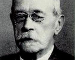
(or Gram's method) is a method of differentiating bacterial species into two large groups (gram-positive and gram-negative). The name comes from its inventor, Hans Christian Gram.
Gram staining differentiates bacteria by the chemical and physical properties of their cell walls by detecting peptidoglycan, which is present in a thick layer in gram-positive bacteria. In a Gram stain test, gram-positive bacteria retain the crystal violet dye, while a counterstain (commonly safranin or fuchsin) added after the crystal violet gives all gram-negative bacteria a red or pink coloring.
♦ Staining mechanism
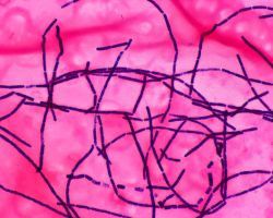
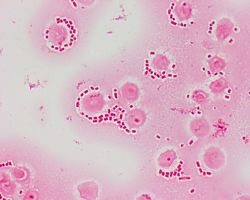
Gram-positive bacteria have a thick mesh-like cell wall made of peptidoglycan (50–90% of cell envelope), and as a result are stained purple by crystal violet, whereas gram-negative bacteria have a thinner layer (10% of cell envelope), so do not retain the purple stain and are counter-stained pink by the Safranin / Fuchsine. There are four basic steps of the Gram stain.
♦ Gram stain
Applying a primary stain (crystal violet) to a heat-fixed smear of a bacterial culture.
Heat fixing kills some bacteria but is mostly used to affix the bacteria to the slide so that they don't rinse out during the staining procedure.
The addition of iodide, which binds to crystal violet and traps it in the cell,
rapid decolorization with alcohol, and
counterstaining with carbol fuchsin.
After decolorization, the gram-positive cell remains purple and the gram-negative cell loses its purple color. Counterstain, which is usually positively charged basic fuchsin, is applied last to give decolorized gram-negative bacteria a pink or red color.
♦ Gram variable
Some bacteria, after staining with the Gram stain, yield a gram-variable pattern: a mix of pink and purple cells are seen.
The genera Actinomyces, Arthobacter, Corynebacterium, Mycobacterium, and Propionibacterium have cell walls particularly sensitive to breakage during cell division, resulting in gram-negative staining of these gram-positive cells. In cultures of Bacillus and Clostridium, a decrease in peptidoglycan thickness during growth coincides with an increase in the number of cells that stain gram-negative. In addition, in all bacteria stained using the Gram stain, the age of the culture may influence the results of the stain.
-
History
History
The method is named after its inventor, the Danish scientist Hans Christian Gram (1853–1938), who developed the technique while working with Carl Friedländer in the morgue of the city hospital in Berlin in 1884. Gram devised his technique not for the purpose of distinguishing one type of bacterium from another but to make bacteria more visible in stained sections of lung tissue. He published his method in 1884, and included in his short report the observation that the Typhus bacillus did not retain the stain.(source:wikipedia)
-
Related
-
References
Wikipedia