Rhodococcus equi (Prescottella equi)
-
General information
R. equi ia a facultative intracellular micro-organism, who can survive an multiply in the macrophages.
Taxonomy
Family: Nocardiaceae
Genus: Prescotella equi
Formerly Corynebacterium equi / Rhodococcus equi
Natural habitats
In the environment, commonly found in dry and dusty soil, domesticated animals, (horses and goats), wild animals, bloodsucking arthropods, plants and in fresh and salt water.
Infection is primarily by inhalation of the bacteria, and also inoculation into a wound or mucous membrane and exposure to domestic animals.
Clinical significance
Are rarely found in humans.
At risk groups are immunocompromised people, such as HIV-AIDS patients or transplant recipients.
R. equi (P. equi) is still the most commonly found of this genus.
They are found in immune compromised patients, such as HIV, in these patients, the infection can be life threatening.
Possible infections, pneumonia, bacteremia, skin infections, endopthalmitis, peritonitis, catheter-related sepsis and abscess of the prostate.
-
Gram stain
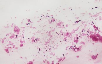
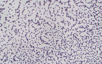
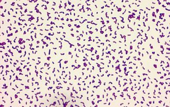
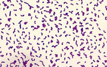
Gram positive rods,
beaded to solidly staining coccobacilli.
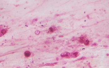
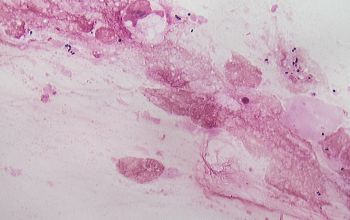
In patient material they are located intracellular in macrophages and extracellular.
They can range from coccoid to bacillary, depending on the stage of growth.
They exhibit a rod-coccus growth cycle
ROD-COC CYCLE
patient material
- pus and tissue: mostly coccoid form visible
- blood, BAL, sputum: usually the rod form visible
liquid medium
rod forms, some branching of individual cells may be found
culture preparations
rod-coc-cycle ranging from rod to coc, depending on the incubation period and growth conditions
- after 4 hours in fluid media 35°C ► rod
- after 24 hours on BA or fluid media 35°C ► cocci
They can easily be dismissed as “diphteroids” because of their gram stain morphology
modified Kinyoun weak
Smear from BA or Choc agar, may appear negative
-
Culture characteristics
-
Rhodococcus grows slowly and is therefore easily overgrown by other flora
Obligate aerobic
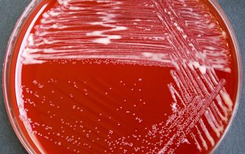
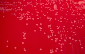
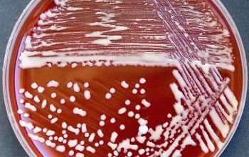
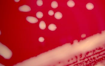
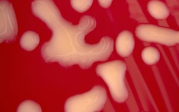
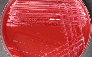
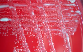
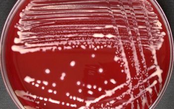
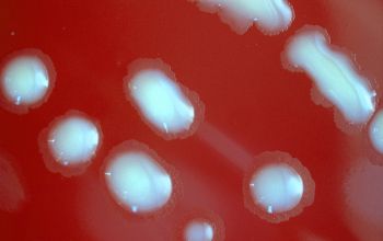
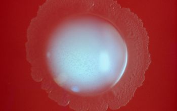
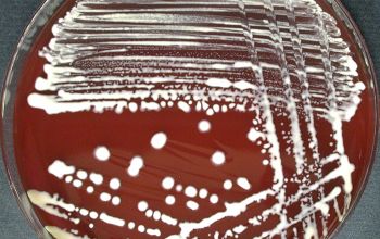
BA: highly variable in their colony morphology, rough, smooth or mucoid and pigmented light yellow, red, orange or dark pink, after a few days of incubation
1 colonies are pale pink and slimy
2 coral red and not slimy (most occurs)
3 pale yellow and not slimy, more opaque than the slimy colony (the least occurrence)
4 colorless colonies
Aerial hyphae: negative
Substrate hyphae: variable
McConkey: growth
BBAØ: no growth
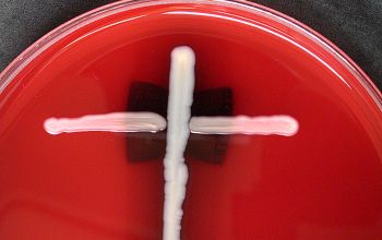
CAMP-test: positive
R. equi (P. equi) is enhanced in the vicinity of the S. aureus streak = CAMP-test positive
-
-
Characteristics
-
References
James Versalovic et al.(2011) Manual of Clinical Microbiology 10th Edition
Karen C. Carrol et al (2019) Manual of Clinical Microbiology, 12th Edition

