Mycobacterium fortuitum (Mycobacteroides fortuitum)
-
General information
M. fortuitum belongs to the family of nontuberculous mycobacteria (NTM) classified in the rapidly growing mycobacteria (RGM)
Taxonomy
Family: Mycobacteriaceae
Genus: Mycobacteroides fortuitum
Formely: Mycobacterium fortuitum
M. fortuitum group
M. fortuitum
M. bonickei, M. houstonense, M. mageritense, M. mucogenicum, M. peregrinum, M. senegalense, M. septicum
Natural habitats / RGM
They are generally considered to be environmental saprophytes which are widely distributed in nature; they have been isolated from soil, dust, natural surface and municipal water, wild and domestic animals, and fish
Contaminated ice machines are a relatively important hospital reservoir for the RGM, especially M. fortuitum
Clinical significance
The most frequently recognized clinical diseases.
Localized post-traumatic wound infections. Surgical wound infections, especially following augmentation mammoplasty, cardiac surgery and catheter infections.
Localized infection in sporadic community-acquired disease usually occurs after a traumatic injury followed by potential soil or water contamination. Such injuries as stepping on a nail, motor vehicle accidents, compound fractures, etc., are typical of the clinical histories seen in patients with RGM disease.
Disseminated infections with M. fortuitum are rare.
The first case report appeared in 1990, when Sack described a patient with a history of intravenous IV drug abuse and AIDS, who had cutaneous lesions from which M. fortuitum was isolated. Cultures of specimens from lymph nodes, urine, pleural effusions, and feces all yielded M. fortuitum.
-
Gram stain
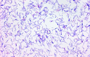
Straight or slightly curved Gram positive rods,
between 0.2-0.6 µm x 1.0-10 µm.
Acid-fast bacilli appear pink in a contrasting background.
Their cell wall has a high lipid content, responsible for acid resistance in Ziehl-Neelsen stain.
They are intracellular organisms resistant to the environmental conditions.
The bacterium is a facultative intracellular parasite, usually of macrophages, and has a slow generation time, 15-20 hours, a physiological characteristic that may contribute to its virulence.
However, microscopy is relatively insensitive, since at least 10,000 organisms per milliliter of sputum are required for smear positivity.
Experienced microscopists may also detect mycobacteria in Gram stained specimens in which they appear as refractile Gram positive or Gram neutral rods.
Under a 100x oil immersion objective, mycobacteria are slightly bent
Cord-factor
Mycobacterium species when grown in a broth medium they may form long cords of cells.
-
Culture characteristics
-
Obligate aerobic
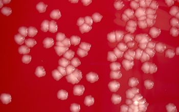
BA: colonies are non-pigmented, rough and wrinkled
Primary isolation within 7 days on multiple types of solid media
and the absence or slow appearance of any pigmentation
Confirmation of the presence or absence of mycobacteria in clinical specimens has traditionally required culture, because of the relative insensitivity of direct microscopy.
In general, clinical specimens that are normally sterile, such as blood, cerebrospinal fluid, or serous fluids, can be inoculated directly onto media. In comparison, nonsterile specimens, such as sputum or pus, must be chemically decontaminated first in order to eliminate common bacteria and fungi that would overwhelm the culture.
However, decontamination procedures may inhibit the growth of mycobacteria, especially NTM, as well.
-
-
Characteristics
-
References
James Versalovic et al.(2011) Manual of Clinical Microbiology 10th Edition
Barbara A. Brown-Elliott Clinical Microbiology Reviews, 2002, vol 5
Microbiology of nontuberculous mycobacteria Author: David E. Griffith UpToDate
Clinical and Taxonomic Status of Pathogenic Nonpigmented or Late Pigmenting Rapidly Growing Mycobacteria


