
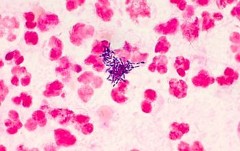
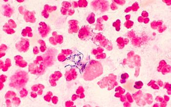
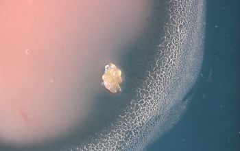
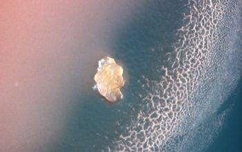
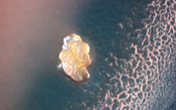
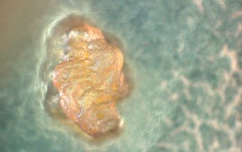
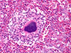
Colony of Actinomyces within an abscess.
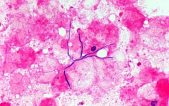
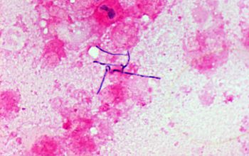
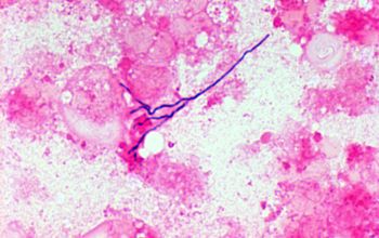
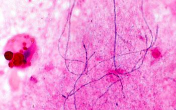
A Gram stain is needed to see the filamentous organisms.
Large colonies of Actinomyces can appear macroscopically as yellow granules whch have been termed "sulfur granules".
The darkly staining eosinophilic rim at the periphery of the colonly consists of immunoglobulin and cell debris which is referred to as the Splendore-Hoeppli phenomenon.
This deposition occurs around colonies of fungi and bacteria and around parasitic organisms
Author: Yale Rosen / Wikimedia Commons
https://commons.wikimedia.org/wiki/File:Actinomycosis_2.jpg
Actinomyces israelii
-
General information
Taxonomy
Family: Actinomycetaceae
Natural habitats
This bacteria is a natural component of the human gastrointestinal and oral flora
Clinical significance
Altough they are not normally a disease causing organism, it is the primary causative agent of Actinomycosis, a long term infection that commonly affects the face and neck.
They form the characteristic aggregates of branching bacilli seen macroscopically as "sulpher granules", there are always multiple polymicrobial 94.5%.
Actinomycosis
The disease is characterised by the formation of painful abscesses in the mouth, lungs or gastrointestinale tract.
Actinomycosis abscesses grow larger as the disease progresses, often over months.
The abscesses often contains characteristic granules (sulphur granules).
-
Diseases
-
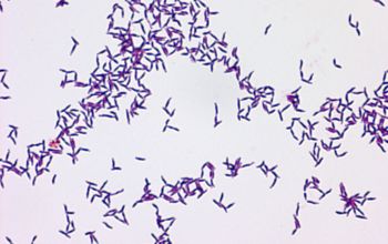
Gram stain
Gram positive branching rods
-
Culture characteristics
-
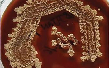
Anaerobic to microaerophilic
They grow better anaerobic than aerobic, or grow poorly or not at all aerobic, but after a number of subcultures they grow aerobically / CO2
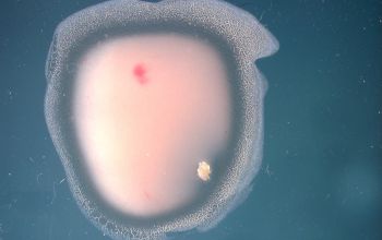
BA: colonies have a rough (“molar tooth”) or smooth surface, not hemolytic
BBA Ø:
- rough colonies: young (2-3 days old) growing like spider shaped colonies they grow a typical shape of a “molar tooth” or “breadcrumb”, often sandy (gritty), hard and stick to the agar (7-14 days), non-hemolytic
- smooth colonies: are slightly elevated, white crème, and opaque
Approximately one third of the strains are smooth and grow faster. In 2-3 days they are 1-2 mm.
Older colonies can become pink
-
-
Characteristics
-
References
James Versalovic et al.(2011) Manual of Clinical Microbiology 10th Edition
Karen C. Carrol et al (2019) Manual of Clinical Microbiology, 12th Edition
Microbiology on the go. An initiative by
Dept. Medical Microbiology and Infectious diseases