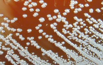
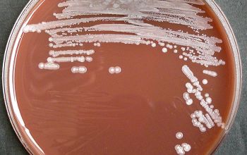
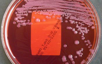
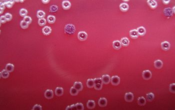
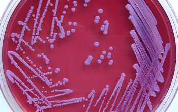
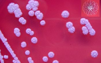
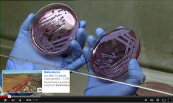
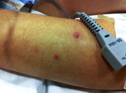
a | Typical colony morphology of B. pseudomallei on Ashdown's agar after incubation at 37°C in air for 3 days. b,c | Colony variation is commonly seen during culture of clinical isolates on Ashdown's agar. (c) shows the variable colony morphology that can be seen from a single sample; genotyping of these colonies showed that one clonal type was present. Colony variation can also be seen within a single colony, as shown in (b) in which the parental colony (pink) has given rise to a second morphotype.
Pictures courtesy of Mrs Vanaporn Wuthiekanun and Mrs Narisara Chantratita, Wellcome Trust, Faculty of Tropical Medicine, Mahidol University, Bangkok, Thailand.
Burkholderia pseudomallei
-
General information
Several Burkholderia species have been isolated from human clinical samples, but only Burkholderia cepacia complex, B. gladiola, B. mallei, B. pseudomallei are generally recognized as human pathogens.
Taxonomy
Family: Burkholderiaceae
Natural habitat
They are found in soil and surface water primarily in tropical and subtropical areas.
They have been isolated in the rice-growing regions of northeast Thailand, western Cambodia, Laos, and southern and central Vietnam. Also in northern Australia, Madagascar and Mauritius.
They can survive deep in the ground and to be rinsed during the rainy season to the surface.
Transmission
There is no person to person transmission
B. pseudomallei is acquired from the environment by inoculation through cut or abraded skin, inhalation, or ingestion.
Clinical significance
B. pseudomallei is the causative agent of the human and animal disease melioidosis.
Melioidosis
is an especially important potential travel related illness for those with CF, and persistent colonization of airways with B. pseudomallei can occur in CF despite prolonged therapy.
Infection with this organism should be considered in the differential diagnosis of any individual with a fever of unknown origin or a tuberculosis-like illness who has a history of travel to a region where B. pseudomallei infection is endemic.
-
Diseases
-
Gram stain
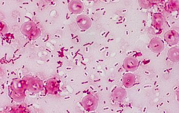
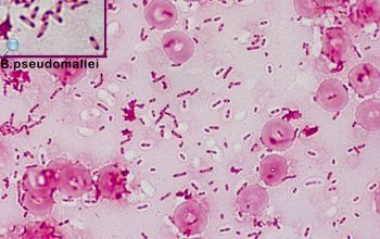
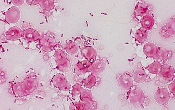
Small Gram negative rods,
0.3 to 1-3 µm,
with bipolar staining, making the cells resemble safety pins.
-
Culture characteristics
-
Obligate aerobic
BA: colonies are typically small, smooth, and gray to pale yellow or creamy in the first 48 hours.
On further incubation, this appearance changes to give dry, wrinkled colonies.
(like Pseudomonas stutzeri)
Colony variation occurs in the same culture.
McConkey: growth
Colorless or pink colonies
Colony variation occurs in the same culture
BBAØ: no growth
Asdown’s agar: (selective agar)
B. pseudomallei colonies on this agar showing the characteristic cornflower head morphology
Smell
musty or earthy odor (don’t sniff on open plates)
-
-
Characteristics
-
References
James Versalovic et al.(2011) Manual of Clinical Microbiology 10th Edition
Karen C. Carrol et al (2019) Manual of Clinical Microbiology, 12th Edition

