SEARCH
Search
Methylene Blue stain
-
General
♦ Methylene Blue stain
Or Loefflers stain is a multipurpose stain recommended for use in staining bacteria and leukocytes..
♦ Summary
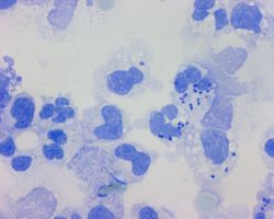
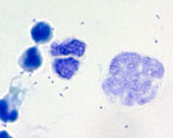
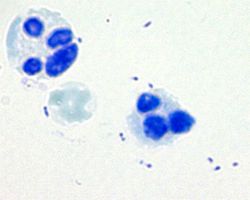
Methylene Blue is recognized as a simple stain used for determining bacterial morphology.
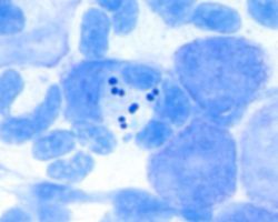
Is recommended for use in the staining of gram-negative bacteria found in spinal fluid, namely Neisseria meningitidis and Haemophilus influenzae.
This tecnique, more so than gram stain, allows for better contrast these gram-negative organisms and the background.
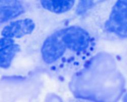
May be used in the presumtive identification of Corynebacterium diphtheria, is due to an accumulation of polymerized polyphosphates in high concentration inside the cell.
This condition appears as polyphosphate granules stained deeply blue, surrounded by lighter blue stained cytoplasm and ar often called Babes-Ernst bodies or metachromatic granules.
The cell itself is characterized by a banded or beaded appearace.
♦ Meningitis
Meningitis of bacterial origin is a severe infection that must receive immidiate attention and prompt treatment.
Thorough initial examination of cerebrospinal fluid (CSF) in the laboratory can yield information which can provide invaluable perspective for proper therapy of patients with this condition.,
Direct microscopic examination of CSF with the Gram stain has proven to be very helpful in the rapid provision of relevant information. Unfortunately, the Gram stain is less sensative with a low number of bacteria from Neisseria meningitidis.
Most studies have reported that only 70 to 85% of culture positive CSF specimens are positive by Gram stain. The lack of contrast staining with gram-negative organisms is another problem with the Gram procedure, particularly if the specimen contains inflammatory cells and proteinaceous material with only a few organisms.
Methylene Blue staining of infected CSF is that it is easy to interpret. Microorganisms stain dark blue, whereas proteinaceous material stain light blue. Much less time is required to detect bacteria with Methylene Blue staining than with Gram staining because of this contrast.
In addition the use of Gram stain to stain material is necessary because gram-negative microorganisms do not stain with the appropiate contrast. It is therefor, necessary to perform an additional stain on positive smears to determine classic Gram stain morphology.
♦ Methylene Blue
Flood the slide with the Methylene Blue. Allow to stand for one to three minutes. Rinse slide with tap water, and blot dry.
Examine the smear at 1000x, using the oil immersion objective.
-
History
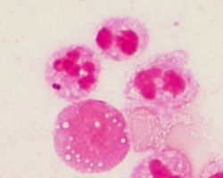
In addition to the staining of microorganisms, Methylene Blue was demonstrated by Harris in 1972 to be an effective stain for the enumaration of leukocytes in mucus of faeces.
-
Related
-
References