


Legionella pneumophila
-
General information
the following information is not yet verified
Taxonomy
Family: Legionellaceae
Natural habitats
Almost all of the Legionella spp have been isolated from aqueous environmental sources.
It is assumed that the natural reservoir of all the Legionellaceae is our aqueous environment and that humans are an accidental host of the bacterium.
Environmental L. pneumophila is a facultative intracellular parasite of several different free-living amoebae, such as Acanthamoeba and Naegleria, existing in microbial consortia in biofilms and free-flowing water.
Many different Legionella spp exist in nature within free-living amoebae, and as a result, these otherwise fastidious bacteria can multiply within the amoebae and be protected from biocides.
Clinical significance
L. pneumophila serogroup 1 causes 95-98% of community-acquired Legionnaires Disease.
Infection is acquired by aerosol inhalation of contaminated water (e.g. cooling towers or aerosol generating devices), although micro aspiration may also be a mechanism of acquiring the disease.
L. pneumophila causing more than 90% of cases of Legionnaires Disease.
Legionnaires Disease,
is a type of bacterial pneumonia, caused by L. pneumophila and other Legionella spp.
The pneumonia ranges in severity from mild to fatal, with an average fatality rate of 12%.
Major risk factors for the disease include immunosuppression of the cellular immune system, cigarette smoking, use of well water, chronic heart or lung disease, and chronic renal failure.
-
Diseases
-
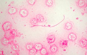
Gram stain
the following information is not yet verified
Lung, sputum
Gram negative rods, 3-5 µm, from small coccobacillus to short rod Culture plate
Gram negative rods, 10-25 µm, usually a long, filamentous bacillus
The small size of intracellular L. pneumophila, they form present in human tissues, makes visualization difficult with Gram stain, as does stain uptake by the surrounding proteinaceous material found in sputum and tissue specimens.
Immunofluorescent microscopy
is the most sensitive and specific microscopic method for the detection of L. pneumophila in tissues and sputum.
-
Culture characteristics
-
the following information is not yet verified
Obligate aerobic
5% CO2 improves the growth
Nutritionally fastidious, growth dependence for L-cysteine and growth enhancement by iron.
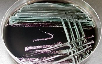
BCYE (buffered charcoal yeast extract)
They grow after 3 days of incubation.
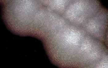
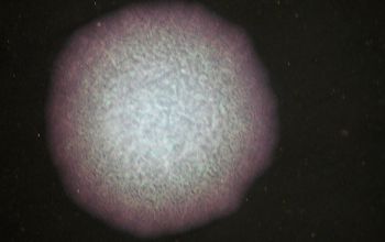
Colonies are flat, entire, and 0.5-1 mm, when observed with a dissecting microscope, they usually have a speckled blue, blue-green, or red color.
By longer incubation, these colonies become smooth, convex, iridescent, and entire and look opal-like when observed with a dissecting microscope.
A thick string may form when a loop is inserted in the colony and then removed from the colony.
BA: no growth
McConkey: no growth
BBAØ: no growth
-
the following information is not yet verified
-
Characteristics
-
References
James Versalovic et al.(2011) Manual of Clinical Microbiology 10th Edition
Karen C. Carrol et al (2019) Manual of Clinical Microbiology, 12th Edition
Microbiology on the go. An initiative by
Dept. Medical Microbiology and Infectious diseases